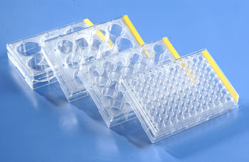
Individualized medicine is one area there is new developments are happening. During the last several years during drug discovery, drug candidates were screened or assays on generic cell lines for the ability to kill these cells, cell binding and any other phenotypic changes. Although the information obtained from these assays were relevant as a whole, it will not have any specific effects to the particular patient being treated. Multiple patients with the same type of cancer, one drug might effect differently for different patients. Some drug will not even respond to certain patients with the same type of cancer, where the drug worked. Although these patients have the same type of cancer, specific cancer types, its microenvironment, its growth pattern, and expression of receptor on the cancer cell surface can be very different from patient to patient.
In the new development, the biopsies of these cancer patients are used to generate individual cells. These cells are then grown in tissue culture flasks or plated in 96 well multiwell plates. Drug compounds are then added directly to the cells at different concentration, and monitor their growth by cell viability or stain for certain marketers by high content image based assay. In this assay, each drug effects at different concentrations specific to that individual patient. That data can be used to fine-tune the therapeutic strategy for that particular patient. This can vary from patient to patient in this type of approach. This method although more labor intensive, will be effective. This approach will also eliminate the drugs, which will not respond to a particular patients cancer cells. In the post-genomic era, this is where the cell-based assay is advancing.
Flow Cytometry
For high content screening, the adherent cells are plated to a 96 well or 384 well cell culture plates. The cells attach to the bottom TC coated surface; an imaging microscope with camera takes the image of the cells at a rapid rate. However for non-adherent cells, this method will not work, because cells will not attach to the bottom surface. There are a number of cancer have non-adherent cells, specifically blood cells related cancer. For this situation, there is a new method called high content flow-cytometry. There are a several instruments are available in the market, where the cells after staining for specific markers are send in a column one by one in a rapid fashion. As the cells pass through a column of medium a high-speed camera take the multiple images of the cells. In some cases up to 5000 cells can be imaged per second, and each cell will have multiple images. Some of the advanced systems, it can differentiate clump of cells rather than individual cells, and can also identify different population of cells in a heterogeneous sample such as blood cells. The images from this screen are saved in a computer and using the software provided, one can do the data mining of the images of specific population of interest or population with different drug treatments. Flow cytometry and high content imaging are two new powerful tools employed in todays drug discovery research.

