High content analysis (HCA) is a versatile tool used in basic research, primary screen for drug discovery efforts where the effect of certain drug compounds are tested on the cells, or target identification, and predicting clinical outcomes. High content analysis is being utilized increasingly in the research and drug discovery screen everyday. Availability of newest technology, wide array of reagents including labeling reagents makes high content analysis very efficient and powerful tool. High content analysis combines three separate tools (1) fluorescent microscopy, which allows the use of wide array of fluorescent labels to label cells and gives flexibility not only to image the cells but also measure cellular changes at different resolution. (2) Ability to quantitate the cellular changes in an automated fashion in a multiwell format using plate readers (3) power of flow cytometry for multi-parametric assessment of specific cell populations. Some of the commonly used applications for HCA is cell signaling, neuronal regeneration, in vitro toxicity, cell migration, angiogenesis, and cell proliferation.
Steps involved in the high content screening
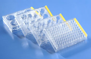
First identify the cells you are interested in high content screening. This could be primary cells or cell lines of interest. If it is primary cells, depending on the cell type, the cell multiwell plates to grow the cells may have to be coated with extracellular matrix protein such as collagen, fibronectin, or laminin. Without the coating appropriate for the cells, they will not attach and grow on the surface of the plate. The ideal coating conditions of the ECM can be obtained from the guidelines from the supplier of the cells. After the cells are identified, plate them in the appropriate tissue culture treated multiwell plates or multiwell plates with grid bottom for easy identification of cells or cell colonies of interest. After the cells are attached, treat them with compounds of interest, or si RNA designed to elicit a response. Next, cells are labeled with fluorescent dye of choice, which targets celluar targets, sub cellular organelles, proteins or cell membrane. Each type of labels work differently based on the cell type. After the necessary incubation with the labels, the cells are imaged using a high content analysis platform. Once the image data from the cells are acquired, the data is analyzed using the software supplied with the instrument for analysis.
Multiplexing
One of the ways to obtain more information from the high content screening is to label multiple cellular targets with different fluorescent labels. Each label will be read at different wavelengths using multichannel fluorescence microscopy, and analyze specific cellular components. When selecting labels, one should take into consideration the problem of overlapping excitation and emission profiles of labels used. As the cost of drug development is steadily increasing, it is important to consider various screening models to reduce the cost. Multiplexed high-content screening is one of the efficient ways to accomplish this.